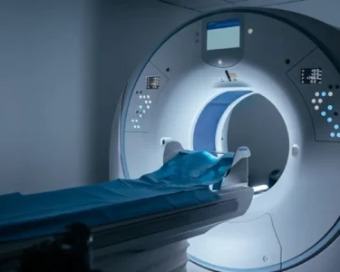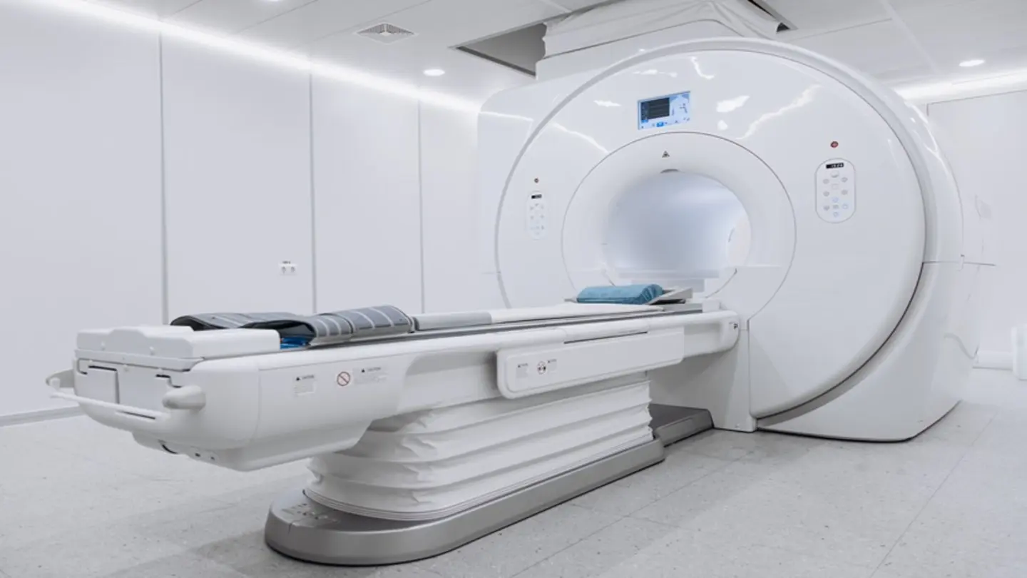
A new study published in the European Medical Journal demonstrates that ultra-high-field 5 Tesla (5T) magnetic resonance imaging (MRI) significantly outperforms the more commonly used 3 Tesla (3T) MRI in the imaging of prostate cancer. The research suggests that the increased magnetic field strength of 5T MRI provides substantially improved image quality, leading to more accurate detection and characterization of cancerous lesions within the prostate.
The study, conducted by researchers at [Information not explicitly provided in the source, but implied to be a European institution], involved a comparison of 5T and 3T MRI scans in a cohort of patients suspected of having prostate cancer. The findings revealed that 5T MRI offered higher resolution images, allowing for clearer visualization of the prostate’s anatomy and the identification of smaller, more subtle tumors that might be missed by 3T MRI.
Improved Detection Rates
Specifically, the 5T MRI demonstrated a greater ability to delineate the peripheral zone of the prostate, a region frequently affected by cancer. This improved delineation is crucial for accurate tumor localization and staging. Researchers noted a statistically significant increase in the detection rate of clinically significant prostate cancer using 5T MRI compared to 3T MRI. This translates to a potentially earlier and more effective diagnosis for patients.
The benefits of 5T MRI extend beyond simply detecting more tumors. The enhanced image quality also aids in differentiating between cancerous and non-cancerous tissue, reducing the likelihood of unnecessary biopsies. Prostate biopsies, while essential for confirming a cancer diagnosis, can be invasive and carry risks of complications such as infection and bleeding. A more accurate imaging technique like 5T MRI could therefore minimize patient discomfort and improve overall care.
While 3T MRI is currently the standard of care for prostate cancer imaging in many healthcare settings, the study highlights the potential for 5T MRI to become a more widely adopted tool. However, the implementation of 5T MRI is not without its challenges. The technology is more expensive than 3T MRI, and requires specialized infrastructure and expertise. Furthermore, the higher field strength can introduce artifacts in the images, requiring careful optimization of scanning protocols.
Despite these challenges, the researchers are optimistic about the future of 5T MRI in prostate cancer diagnosis. They believe that ongoing advancements in technology and image processing techniques will further enhance the capabilities of 5T MRI and make it more accessible to patients. The study’s authors suggest that wider availability of 5T MRI could lead to a paradigm shift in prostate cancer management, enabling more precise diagnosis, personalized treatment planning, and ultimately, improved patient outcomes. Further research is planned to investigate the long-term impact of 5T MRI on prostate cancer survival rates.
The findings underscore the importance of continued investment in advanced imaging technologies to improve the early detection and treatment of cancer. The potential benefits of 5T MRI for prostate cancer patients are substantial, and warrant further exploration and clinical validation.
Image Source: Google | Image Credit: Respective Owner