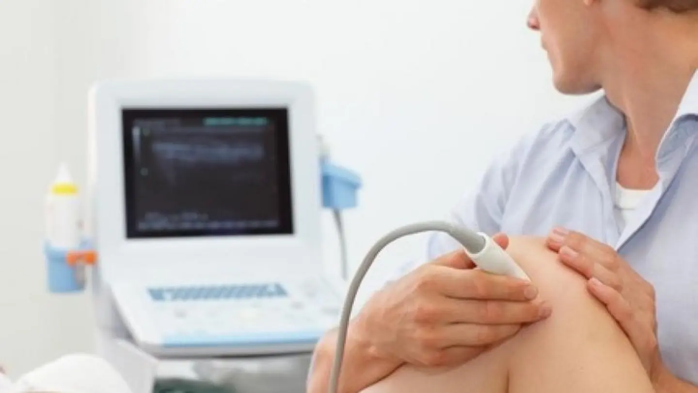
Point-of-care ultrasound (POCUS) has become a transformative tool in rheumatology, replacing the traditional stethoscope’s auditory limitations with real-time visualisation. Unlike stethoscopes, POCUS reveals synovial effusion, soft tissue abnormalities, and fluid collections immediately, offering insights that alter patient management trajectories in inflammatory conditions. This technology democratises access to advanced diagnostics, especially in resource-limited settings.
POCUS monitors treatment response via serial scans tracking synovial volume and vascularity changes. Objective metrics correlate with clinical improvement, invaluable in chronic diseases like rheumatoid arthritis where timely assessment prevents flare-ups. It reduces costs by ruling out complex conditions.
POCUS enhances patient-physician communication. Dynamic imaging translates abstract clinical descriptors into tangible visuals, improving understanding and treatment adherence. Studies show patients receiving ultrasound-guided explanations report higher satisfaction and comprehension of disease activity.
The adoption curve reflects confidence in training programs. Modern residencies mandate ultrasound competency, recognising it as essential for comprehensive care. Challenges persist: interpretation variability, need for standardised protocols, and potential over-reliance on imaging without clinical context.
AI integration promises to streamline image analysis, reducing learning curves and improving accuracy. As POCUS evolves, it will become the definitive rheumatology assessment tool—providing rapid, non-invasive, radiation-free evaluation empowering clinicians and patients.
The article underscores POCUS applications in arthritis and lupus. In arthritis, it detects subclinical synovitis and bone erosions, enabling pre-emptive treatment adjustments. For lupus, ultrasound identifies tenosynovitis and cutaneous manifestations. It differentiates inflammatory from mechanical pain, reducing misdiagnosis.
POCUS enhances patient-physician communication. Dynamic imaging turns abstract clinical descriptors into tangible visuals, fostering better understanding and adherence to treatment plans. Studies cited demonstrate higher satisfaction when patients receive ultrasound-guided explanations of disease activity.
The European Medical Journal editorial stresses structured training to maximise POCUS utility. Formal certification ensures consistent image acquisition and interpretation. EULAR guidelines recommend routine ultrasound for high-sensitivity conditions, integrating it into standard practice protocols.
The authors caution against over-dependence. “POCUS complements, not replaces, clinical evaluation,” notes Dr. Elena Martinez. Imaging must be interpreted alongside patient history and lab data. Otherwise, clinicians risk missing subtle clues only holistic assessment reveals.
Future innovations include AI-enhanced portable devices that flag pathology autonomously. EHR integration enables longitudinal tracking of joint changes, offering predictive insights for personalised medicine. As healthcare prioritises value-based care, POCUS stands out as a low-cost, high-impact intervention enhancing diagnostic precision and patient experience.
Image Source: Google | Image Credit: Respective Owner