Metabolic-associated steatotic liver disease (MASLD) has become a critical health concern globally, driving the need for precise diagnostic tools. Liver biopsy remains a cornerstone for assessing the disease’s progression and guiding treatment. However, recent studies highlight its limitations, including invasive nature and variable accuracy in detecting early-stage lesions. This article explores the efficacy of liver biopsy in MASLD diagnosis, the obstacles clinicians face, and emerging alternatives.
Role of Liver Biopsy in Diagnosis
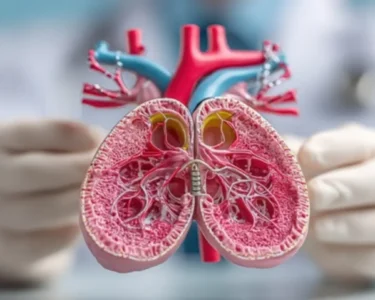
Traditionally, liver biopsy is considered the gold standard for evaluating liver fibrosis and steatosis in MASLD. It involves extracting a small tissue sample for histopathological analysis. While this method provides detailed insights into disease severity, its reliability is often compromised by sampling error. Studies show that up to 30% of biopsies may underestimate fibrosis, leading to delayed interventions. Additionally, the procedure carries risks like bleeding or organ damage, particularly in obese patients with comorbid conditions.
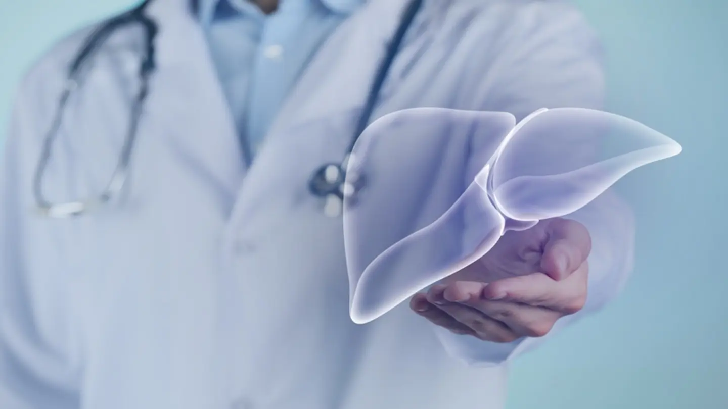
Diagnostic accuracy hinges on the skill of the hepatologist performing the biopsy. Variability in technique and interpretation can skew results. For instance, distinguishing between fibrous and fatty tissue is challenging, especially in cases of overlapping pathologies. This inconsistency underscores the need for complementary non-invasive methods to validate biopsy findings.
Challenges in Clinical Practice
One major hurdle is patient compliance. Many individuals delay or refuse biopsies due to fear or discomfort. This reluctance is exacerbated by the fact that early-stage MASLD is often asymptomatic. Clinicians must balance the diagnostic benefits of biopsy against patient willingness. Moreover, the cost and time associated with biopsies, including hospitalization in some cases, add to the burden on healthcare systems.
Technical challenges further limit biopsy reliability. Obesity, a common feature of MASLD, makes tissue sampling technically difficult. The thicker abdominal wall and higher fat content can obscure the liver, increasing the risk of improper sampling. These factors collectively contribute to diagnostic uncertainty, prompting researchers to seek alternatives.
Alternatives to liver biopsy, such as transient elastography and FibroScan, offer non-invasive imaging of liver stiffness. However, these methods lack the specificity needed to distinguish MASLD from other conditions like viral hepatitis. Advances in blood-based biomarkers, including ALP and GGT levels, show promise but are still under validation. Artificial intelligence is also being explored to improve imaging interpretation, though integration into clinical practice remains limited.
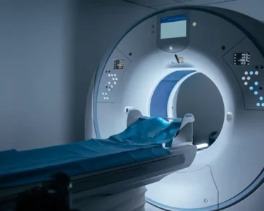
Despite these advancements, liver biopsy retains value in complex cases where non-invasive tests yield ambiguous results. Ongoing research aims to refine biopsy protocols, such as targeted biopsy using CT or MRI guidance, to enhance accuracy. Standardizing diagnostic criteria could also mitigate variability in interpretation.
In conclusion, while liver biopsy is indispensable in MASLD management, its drawbacks highlight the urgency for hybrid diagnostic approaches. A combination of biopsy with high-resolution imaging or biomarker panels may offer a more reliable pathway to early detection and personalized treatment.
Image Source: Google | Image Credit: Respective Owner